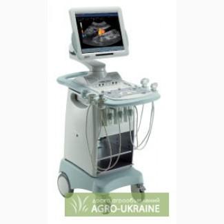
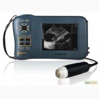
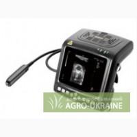
/ Veterinary equipment, preparations / Veterinary equipment / Veterinary ultrasound apparatus MyLab40Vet
For sale / buy
Veterinary ultrasound device MyLab40Vet, Poltava region.
Region :
Poltava region.
Updated:
MyLab40Vet - universal ultrasound system for veterinary research , an ideal choice for veterinary clinics and large horse breeding farms.
Full range of ultrasonic modes and modern technologies:
The MyLab40Vet ultrasound device is based on the MyLab World modular architecture, which allows you to use a full range of ultrasound modes, innovative packages and technologies. MyLab40Vet can be equipped with modules for cardiology (the specialized cardiology package includes Compass M mode and tissue doppler (Tissue Velocity Mapping)), a module for visualization of reproductive organs of animals and contrast ultrasound imaging. The use of modern technologies and ultrasound modes makes it possible to significantly expand the informativeness and diagnostic accuracy of screening and specialized examinations in veterinary medicine.
A wide selection of ultrasonic sensors:
The developers of the MyLab40Vet ultrasound device took into account all the needs of modern veterinary medicine. The ultrasound system has 3 connectors for connecting broadband phased, convex and linear sensors with a frequency of up to 18 MHz, as well as a Doppler sensor. The spectrum of used sensors includes linear endorectal sensors, which are necessary for the study of reproductive function incattle and horses.
Specialized veterinary software package:
Software package 9.10 implemented in MyLab40Vet includes calculations of abdominal organs of dogs, cats and horses. In the gray-scale mode, it is possible to measure 14 main abdominal organs - kidneys, urinary system, adrenal glands, pancreas, spleen, gastrointestinal system, lymphatic system, liver, gall bladder, prostate, uterus, ovary, upper vena cava cava) and portal veins (Portal Vein).
In Doppler modes, 4 groups of measurements are possible: hepatic veins, renal artery, portal veins, hepatic artery. Measurements of the speed of systole and diastole, as well as the resistance index of the renal and hepatic arteries.
Optimal communication system for veterinary clinics:
Communication in MyLab40Vet is represented by USB ports, a CD/DVD drive and a modern MyLab Desk data management system. The obtained image can be saved in various formats (BMP, JPEG, movie clips) and when choosing the final format, the operator can easily save the research data to the integrated hard disk, burn it to a DVD/CD disk, USB drive or other digital media. It is possible to connect the printer via USB or PC connection, as well as transfer data via a wireless network.
The MyLab Desk software package is an ideal solution for private veterinary clinics and farms to improve the quality and productivity of work. MyLab Desk runs the MyLab40Vet interface on a standard personal computer and allows you to conduct a more detailed study of the obtained results. MyLab Desk is a modern and ideal way of communication between MyLab40 Vet and a personal computer.
Efficiency through design and ergonomics:
The grayscale and color ultrasound picture is displayed on a 19" LCD monitor, which is mounted on a hinged bracket, which increases the comfort of the examination and gives the best quality of the ultrasound picture. The rational MyLab40Vet console allows you to additionally equip the ultrasound system with a printer, as well as to easily move it from department to department. Construction The keyboard of the ultrasonic device is ergonomic and intuitive. All the above factors significantly increase the efficiency and quality of the operator's work Features of the ultrasonic device:
Характеристики ультразвукового аппарата:
Scanning methods: linear, convex, microconvex, phased.
Image acquisition modes: B mode, color B mode, M mode, anatomical M mode, PW/CW mode, СFM mode, Power Doppler, tissue Doppler mode
Combinations of screen modes: orientation - right/left, up/down, triplex model in real time mode. All combinations in color
Connector for simultaneous connection of 3 sensors (convex, microconvex, linear and phased sensors) and convector for connecting a Doppler sensor
Fully digital signal processing in all modes
Visualization in B mode: frequency depends on the technical characteristics of the sensor, depth 4-27 cm, 256 shades of gray, pulse repetition frequency 2.6 - 11 kHz, biopsy line
Visualization in M mode: possible with all sensors, depth 4 - 27 cm, scanning time 2 - 12 seconds, pulse repetition frequency 1 kHz, acoustic zoom, time markers
Visualization in CFM mode: frequency 2.0 - 5.0 MHz, pulse repetition frequency (PRF) 2600 - 33300 Hz, sensitivity level adjustment, 3-level wall filter (Wall filter)
Visualization in SW/PW mode: frequency 2.0 - 5.0 MHz, pulse repetition frequency (PRF) 2600 - 33300 Hz (in PW mode), scanning time 2 - 12 seconds, wall filter (Wall filter) 50 - 1800 Hz, 9 degrees of rejection, volume sample size 1-24 mm (in PW mode), controllable radiation angle 20 degrees (in PW mode), angle correlation +/- 75 degrees, maximum blood flow speed (in SW mode) 6.42 m/s at 2.0 MHz
Scaling of images (Zoom) x 32
The Zoom function is effective in real-time mode, as well as in Freeze mode.
Computational packages for veterinary studies of dogs, cats and horses
Schematic images of anatomical parts of the body so-called body marks
TEI (Enhanced Tissue Imaging) – the use of second harmonic technology, which is supported by all types of sensors
XView - interactive adaptive image processing algorithm
M-View - combined standard and multidirectional ultrasound imaging for detection of all anatomical structures
Universal package of Dicom programs for creating, archiving and transferring research results (Dicom Media Storage, Dicom Store SCU, Dicom Print, Dicom Worklist, Dicom MPPS, Dicom Storage commitment, Dicom Echocardio Structure Report
Ability to install the latest licenses Stress Echo, Contrast(CnTI), Panoramic View (VPan), 3D/4D, X - strain and also Commpass M mode
19'' TFT monitor
Digital equipment: Pentium III 800 MHz processor, 256 MB RAM, Win XP operating system, installed Dicom service classes and data storage, 20 GB hard disk, CD-ROM drive (DVD-RW)
Built-in QWERTY/YTSUKEN keyboard
Connecting ports - RS – 232, LAN RJ45, 2 USB(//tractor-service.com)
Dedicated ports – audio in/out, ECG port, footswitch port, additional port
Video input - output - XVGA output, SVHS output/input, VHS input, RGB, video standart PAL/NTSC
Storage of research results in WMP, PNG, JPG, AVI format
Power consumption 200 W with monitor
Weight without monitor - 60 kg.
Used sensors:
CA621 – a convex sensor with a frequency of 2-7 MHz with a radius of curvature of 60 mm for abdominal studies of large animals
CA631 – a convex sensor with a frequency of 1-8 MHz with a radius of curvature of 60 mm for abdominal studies of large animals
CA431 - a convex sensor with a frequency of 1-8 MHz with a radius of curvature of 40 mm for abdominal studies of large animals.
CA430E - convex sensor with a frequency of 1-5 MHz with a radius of curvature of 40 mm for abdominal studies of large animals
CA1421 – a convex sensor with a frequency of 2-6 MHz with a radius of curvature of 40 mm for abdominal studies of large animals
CA421 – a convex sensor with a frequency of 2-5 MHz with a radius of curvature of 40 mm for abdominal studies of large animals
CA123 - microconvex sensor with a frequency of 3-9 MHz with a radius of curvature of 14 mm for abdominal examinations of domestic animals
C5-2 R13 microconvex sensor with a frequency of 2-5 MHz with a radius of curvature of 10 mm for abdominal studies of large animals
L532E - a linear sensor with a frequency of 2 - 9 MHz for studies of small and medium animals, studies of joints and tendons in horses
L523 - a linear sensor with a frequency of 4 - 14 MHz for the study of small and medium-sized animals, studies of joints and tendons in horses
L522E – broadband linear sensor with a frequency of 2-9 MHz for the study of joints and tendons in horses
L435 - a linear sensor with a frequency of 6 - 18 MHz for studies of small and medium animals, studies of joints and tendons in horses, as well as studies in the field of veterinary ophthalmology
LV513 – linear sensor 5 – 10 MHz for studying the reproductive function of large animals, studying joints and tendons in horses
PA230E – phased sensor with a frequency of 1-4 MHz for abdominal and cardiac studies of large animals
PA122E is a 3-8 MHz phasing sensor for research in the field of veterinary cardiology
PA121E – phased sensor with a frequency of 2-5 MHz for research in the field of veterinary cardiology
PA023E – phasing sensor with a frequency of 5-10 MHz for research in the field of veterinary cardiology of small animals
IOE323 – a linear specialized sensor with a frequency of 4-13 MHz for studies of the reproductive function of large animals and during laparoscopic interventions
LP323 - a specialized linear sensor with a frequency of 4-13 MHz for laparoscopic interventions
TEE022 – a specialized transesophageal phased array sensor with a frequency of 3-7.5 MHz for research in the field of veterinary cardiology
2CW – Doppler sensor with a frequency of 2 MHz
5CW - Doppler sensor with a frequency of 5 MHz
|
Author, contacts | |
ЧП Медикос/ отзывы , info. / activity assessment | |
|
Phone:
0956xxxxxx
show
| |
| http://www.medikos.com.ua | |
Ad ID: #153150
(added by a registered user, registration date: 12-15-2012)
Added / Updated: 2024-08-29 11:05 (relevant, until: 29-08-2025)
Permanent ad address:
Showed / watched for today: ?, total: ?
Similar ads
Among them are many interesting...


